Prevention and Screening: Major Contributors to Saving Lives in Five Key Cancer Types

A recent study led by the National Institutes of Health (NIH) and published in JAMA Oncology highlights the significant impact of prevention and screening in reducing mortality from five major cancer types over the last 45 years. The research focused on breast, cervical, colorectal, lung, and prostate cancers, which collectively represent nearly half of all cancer diagnoses and deaths. This study underscores that while treatment advances are often viewed as the primary contributors to mortality reduction, prevention and screening have played a more substantial role in saving lives.

Co-lead investigator Katrina A. B. Goddard, Ph.D., from the NCI’s Division of Cancer Control and Population Sciences, emphasized that 80% of the deaths averted from these cancers since 1975 can be attributed to advances in prevention and screening rather than treatment. Particularly notable is the impact of smoking cessation, which alone accounted for 3.45 million lives saved from lung cancer. The study further found that prevention and screening were the predominant factors in reducing deaths from cervical, colorectal, lung, and prostate cancers, while treatment advances led in breast cancer mortality reduction.

The study utilized statistical models to analyze cancer mortality data between 1975 and 2020, revealing that 5.94 million deaths were prevented due to interventions in these five cancers, with prevention and screening accounting for 4.75 million of these. The breakdown by cancer type showed varying contributions of screening and treatment. For example, in lung cancer, tobacco control was overwhelmingly effective, accounting for 98% of the averted deaths. Conversely, in breast cancer, treatment was the major contributor, although mammography screening also played a significant role.

These findings underscore the necessity of a multifaceted approach to cancer reduction, combining effective prevention, screening, and treatment strategies. W. Kimryn Rathmell, M.D., Ph.D., director of NCI, stated that understanding which strategies are most effective helps guide future efforts to reduce cancer mortality further. The study also points to recent advancements like HPV vaccination and lung cancer screening, which could potentially enhance these effects even though they were not widely used during the study period.

However, the researchers acknowledge that the study’s focus on these five cancers, which represent less than half of all cancer deaths, means the findings might not apply to cancers without effective prevention, screening, or treatment options. Dr. Philip E. Castle, another co-lead investigator, emphasized the importance of optimizing and expanding access to proven interventions, particularly for underserved populations, and developing new strategies for other lethal cancers such as pancreatic and ovarian cancer.

The authors also noted limitations, including the study’s reliance on population averages and its exclusion of potential screening harms like false positives and overdiagnosis. These findings present a complex picture of cancer prevention and treatment, highlighting the critical role of integrated strategies in ongoing efforts to reduce cancer mortality across various populations.


______________

RadParts, a TTG Imaging Solutions Company, is the world’s largest independent distributor of OEM replacement parts. We specialize in low-cost parts for repairing linear accelerators and radiation equipment. Our mission is to provide high-quality, user-friendly, low-cost components and support for linear accelerators and radiation equipment. Contact RadParts at 877-704-3838 to learn more.


Written by the Digital Marketing Team at Creative Programs & Systems: https://www.cpsmi.com/.
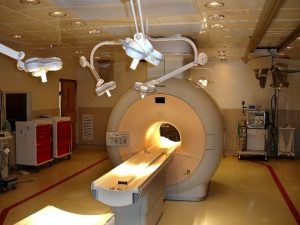
 

Can AI Improve Early Detection of Breast Cancer in MRI Studies?

Recent research indicates that using artificial intelligence (AI) to reevaluate breast MRIs deemed high-risk could markedly enhance early breast cancer detection. A retrospective study published in Academic Radiology examined the effectiveness of a convolutional neural network trained on breast MRI data to predict breast cancer development within a year following initially negative MRI results. The study involved 3,029 MRI scans from 910 patients (average age 52), including 115 cases where breast cancer was later diagnosed on MRI. Initially, all patients received a BI-RADS assessment of less than 3.

The study found that the AI model predicted breast cancer within one year with a 72 percent accuracy as measured by the area under the receiver operating characteristic curve (AUC). Of the initial MRI scans, 83 out of 115 cases showed visual correlations with biopsy-proven breast cancer, with the majority of these correlations measuring less than 0.5 cm.

The researchers emphasized that using the AI model to reassess MRIs identified as high-risk could improve early breast cancer detection rates by up to 30 percent. “It’s crucial to recognize that the reported 30% sensitivity refers to cancers that would likely have remained undetected until the subsequent exam. These are additional detections, supplementing those already identified with high sensitivity by radiologists,” explained lead study author Dr. Lukas Hirsch of the City College of New York.

The study also highlighted that with the AI model’s reassessment, the positive predictive value (PPV) could reach 6 percent for high-risk MRI scans. If radiologists were to recall only half of these cases, they would achieve the PPV standard for tissue diagnosis and detect at least an additional 15% of tumors, representing a clinically significant improvement.

Furthermore, the AI model successfully pinpointed the future location of breast cancer in 57 percent of the cases, with a 71 percent accuracy rate in the 35 true positive cases.

Despite the promising results, the study’s authors acknowledged several limitations, including the single-center, retrospective nature of the study, the assessment limited to sagittal scans, and the small number of cancers detected through screening.

_________

RadParts, a TTG Imaging Solutions Company, is the world’s largest independent distributor of OEM replacement parts. We specialize in low-cost parts for repairing linear accelerators and radiation equipment. Our mission is to provide high-quality, user-friendly, low-cost components and support for linear accelerators and radiation equipment. Contact RadParts at 877-704-3838 to learn more.

Written by the Digital Marketing Team at Creative Programs & Systems: https://www.cpsmi.com/

BAT: A Revolutionary Garment Enhancing Radiation Safety for Healthcare Professionals

On September 23, 2024, Burlington Medical announced the availability of BAT, a groundbreaking radiation protection garment designed to significantly reduce the risk of breast cancer and other radiation-associated cancers among healthcare workers. This innovative garment covers crucial areas such as the breast, axilla, and thyroid.

Developed by Dr. Lauren Ramsey, a Breast Surgical Oncologist, BAT was inspired by a personal loss. Dr. Ramsey’s friend and colleague succumbed to breast cancer, which she believed was exacerbated by occupational radiation exposure. Addressing a dire need within the medical community, BAT offers an additional layer of protection that is often missed by traditional radioprotective aprons.

Dr. Ramsey highlights the increased risk faced by female healthcare professionals, particularly noting, “Female orthopedic surgeons have up to a three times higher risk of developing breast cancer.” With women now comprising 55 percent of medical school attendees, the design of BAT is tailored specifically with their safety in mind. It achieves over 97 percent reduction in radiation exposure to the upper outer quadrant of the breast and also lessens exposure to the thyroid, chest, and spine.

Key benefits of BAT include:

• Targeted Protection: BAT is meticulously crafted to shield the breast, axilla, and thyroid, cutting down radiation exposure by up to 97.7 percent.

• Comfort and Mobility: Featuring a lightweight design with mesh panels, BAT ensures comfort, freedom of movement, and temperature control.

• Compatibility: Designed to be worn with any radioprotective apron, BAT requires no additional attachments, making it a versatile and essential addition to any healthcare professional’s safety gear.

• Enhanced Safety: Besides safeguarding the breast and axilla, it also minimizes scatter radiation that can impact the chest, neck, and spine.

Exclusively distributed by Burlington Medical, BAT stands out as a vital innovation in the field of healthcare safety, offering a critical solution to reduce cancer risks associated with ionizing radiation exposure, which accumulates over time. This garment empowers healthcare workers by providing effective protection without compromising on comfort or mobility.

Healthcare professionals can directly place orders at: https://burmed.com/store/bat.html

___________

RadParts, a TTG Imaging Solutions Company, is the world’s largest independent distributor of OEM replacement parts. We specialize in low-cost parts for repairing linear accelerators and radiation equipment. Our mission is to provide high-quality, user-friendly, low-cost components and support for linear accelerators and radiation equipment. Contact RadParts at 877-704-3838 to learn more.

Written by the digital marketing team at Creative Programs & Systems: https://www.cpsmi.com/

Research Advocates for Advanced Precision Radiation Therapy in Lung Cancer

A new study from The University of Texas MD Anderson Cancer Center advocates for the use of intensity-modulated radiotherapy (IMRT) over 3D-conformal radiotherapy (3D-CRT) in treating unresectable, locally advanced non-small cell lung cancer (NSCLC). Published in JAMA Oncology, the study reveals that IMRT leads to fewer severe side effects, such as pneumonitis, while delivering similar survival outcomes compared to 3D-CRT. This finding is based on a secondary analysis of long-term data from 483 patients enrolled in the Phase III NRG Oncology-RTOG 0617 trial.

The research highlights the technological advancements in IMRT, which unlike the older 3D-CRT that uses straight radiation beams, employs complex computational methods to modulate radiation beams, allowing for a more precise targeting of tumors and sparing of normal tissue. Despite concerns about the long-term effects of low-dose radiation exposure from IMRT, the study confirms that this does not lead to increased secondary cancers or toxicity over time. This makes IMRT a preferable choice in the management of NSCLC, noted for its precision over the rudimentary 3D-CRT, which has been in use for over 50 years.

Statistical analysis of the study data shows that patients treated with IMRT had slightly better, though not statistically significant, five-year overall and progression-free survival rates compared to those treated with 3D-CRT. Even patients with larger or unfavorably located tumors near the heart showed improved outcomes with IMRT. The study emphasizes the need to minimize cardiac exposure during radiation planning, linking lower heart exposure to better median survival rates.

Endorsed by lead author Stephen Chun, the study supports a shift towards prioritizing IMRT in lung cancer treatment, similar to its adoption in other cancers like prostate and brain tumors. This shift reflects a broader move towards maximizing treatment precision to improve patient outcomes while reducing side effects. The research was supported by the National Institutes of Health and other organizations, reinforcing its findings within the oncological community.

For top-quality, user-friendly, and affordable parts support for linear accelerators and radiation equipment, reach out to the experts at RadParts.

RadParts, a TTG Imaging Solutions Company, is the world’s largest independent distributor of OEM replacement parts. We specialize in low-cost parts for repairing linear accelerators and radiation equipment. Our mission is to provide high-quality, user-friendly, low-cost components and support for linear accelerators and radiation equipment. Contact RadParts at 877-704-3838 to learn more.

Written by the digital marketing team at Creative Programs & Systems: www.cpsmi.com

Advancements in Lung Cancer Detection and Treatment Monitoring

UC Davis Health has implemented two groundbreaking imaging technologies to improve lung cancer care. The first technology, a new mobile 3D CT imaging system, enhances early detection of lung cancer by enabling more precise biopsies. This system, used in conjunction with a robotic-assisted bronchoscopy tool, allows for accurate real-time imaging, reducing procedure times and increasing safety. The results have shown a significant increase in early-stage lung cancer diagnoses, which are more treatable.

The second technology involves advanced imaging to monitor the effectiveness of lung cancer treatments. Utilizing the novel EXPLORER PET scanner, researchers can now better understand the lung’s unique blood supply, which helps in evaluating how lung tumors respond to treatments. This technology promises to refine therapeutic strategies, particularly in how treatments such as immunotherapy are applied.

These innovations not only offer hope for early detection but also provide critical insights into treatment efficacy, potentially transforming lung cancer management and outcomes.

For parts and services related to Linear Accelerators and more, contact RadParts!

RadParts, a TTG Imaging Solutions Company, is the world’s largest independent distributor of OEM replacement parts. We specialize in low-cost parts for repairing linear accelerators and radiation equipment. Our mission is to provide high-quality, user-friendly, low-cost components and support for linear accelerators and radiation equipment. Contact Radparts at 877-704-3838 to learn more.

Written by the digital marketing team at Creative Programs & Systems: www.cpsmi.com

Diagnosing Multiple Sclerosis (MS)


Picture via Healthline

In the United States, nearly one million people live with multiple sclerosis (MS), an immune-mediated inflammatory disease that affects the central nervous system by causing demyelination and axonal transection, leading to regional and whole-brain atrophy. Characterized by symptomatic episodes that can occur months or years apart, MS symptoms vary widely but commonly include sensory loss, spasticity, bladder and sexual dysfunction, fatigue, and muscle weakness.

Key Points in MS Diagnosis:

Prevalence and Impact: MS is a major neurological cause of disability among young adults, often diagnosed between the ages of 20 and 40. Although traditionally considered more common among young White individuals, recent data suggest a higher incidence in Black populations. Women are diagnosed about three times more often than men, similar to other autoimmune diseases. The disease significantly affects quality of life, primarily due to physical disabilities and associated conditions like depression and anxiety.

Diagnostic Criteria: MS is diagnosed based on clinical signs and the McDonald Criteria, which require a clinical event indicative of demyelination, such as optic neuritis, brainstem syndrome, or transverse myelitis. These events must present symptoms consistent with the disease for at least 24 hours, supported by neurological exams that point to central nervous system involvement.

Imaging and Tests: MRI is crucial for MS diagnosis but must be interpreted by experts to differentiate MS from other conditions like migraines or ischemic changes. Other tests include cerebrospinal fluid analysis and blood tests to exclude diseases with similar symptoms.

Cognitive and Physical Symptoms: Cognitive impairment, affecting 40%-65% of MS patients, includes problems with memory, concentration, and multitasking, often occurring early in the disease. Recognizing and managing these symptoms, along with physical ones like spasticity and fatigue, is vital for improving patient quality of life.

Advancements in Treatment: MS treatment has evolved significantly, especially with the introduction of B cell-depleting therapies in 2017, which have shifted understanding and management of the disease. Current treatments offer various administration methods, including oral and infusion options, and continue to improve with ongoing research into new therapies like tolebrutinib.

Understanding MS involves recognizing its complex symptoms and staying informed about the latest diagnostic and treatment strategies to manage and mitigate its impact effectively.

_____________

RadParts, a TTG Imaging Solutions Company, is the world’s largest independent distributor of OEM replacement parts. We specialize in low-cost parts for repairing linear accelerators and radiation equipment. Our mission is to provide high-quality, user-friendly, low-cost components and support for linear accelerators and radiation equipment. Contact RadParts at 877-704-3838 to learn more.

Written by the digital marketing team at Creative Programs & Systems: www.cpsmi.com

Researchers Have Created a Perovskite X-ray Detector for Medical Imaging Applications

Image taken from ScienceDirect.com

Researchers from the Shenzhen Institute of Advanced Technology (SIAT) of the Chinese Academy of Sciences and Central China Normal University have developed a high-performance perovskite X-ray detector, designed to enhance medical imaging. Published in Nature Communications, the study introduces a direct-conversion X-ray detector that integrates a thick perovskite film with a CMOS pixel array, offering improved spatial resolution and lower radiation doses.


Traditional semiconductor materials used in X-ray imaging, like silicon and cadmium zinc telluride, often suffer from low X-ray absorption efficiency or high costs. The newly developed perovskite detector addresses these issues by using a CsPbBr3 perovskite film, which demonstrates high detection sensitivity and a significantly lower dose detection limit.


The detector achieves excellent spatial resolution and has been successfully tested in 2D and 3D CT imaging, even at high speeds of 300 frames per second. This advancement indicates a promising future for medical imaging technologies, making X-ray procedures safer and more effective.

________________

RadParts, a TTG Imaging Solutions Company, is the world’s largest independent distributor of OEM replacement parts. We specialize in low-cost parts for repairing linear accelerators and radiation equipment. Our mission is to provide high-quality, user-friendly, low-cost components and support for linear accelerators and radiation equipment. Contact RadParts at 877-704-3838 to learn more.

Written by the digital marketing team at Creative Programs & Systems: www.cpsmi.com

Study Reveals Oncologists’ Perspectives on AI Ethics in Cancer Care

As artificial intelligence (AI) becomes more prevalent in cancer care, oncologists are encountering new ethical dilemmas. Currently, AI is mainly utilized as a diagnostic aid, helping to detect tumor cells in pathology slides and identify tumors in X-rays and other radiological images. However, emerging AI models that can evaluate a patient’s prognosis and potentially suggest treatment options are under development. This advancement raises significant legal questions regarding accountability if an AI-suggested treatment leads to adverse outcomes for a patient.

A recent survey conducted by Dana-Farber Cancer Institute and published in JAMA Network Open on March 28, 2024, reveals insights from over 200 U.S. oncologists on the responsible use of AI in their field. Key findings of this study include:


• Understanding and Consent: 85% of oncologists believe they should understand how AI models function, yet only 23% think patients need the same depth of understanding. However, 81% agree that patient consent is essential when AI aids treatment decisions.


• Decision-Making: When AI suggests a different treatment than the oncologist, 37% would offer both choices to the patient, highlighting the value of shared decision-making.


• Responsibility: A significant 91% of oncologists think AI developers should be accountable for medical or legal issues arising from AI use, far exceeding those who believe responsibility should also involve physicians (47%) or hospitals (43%).


• Bias Protection: While 76% of respondents recognize the need to shield patients from biased AI tools, only 28% feel confident in their ability to detect such biases.


The study underscores the importance of involving oncologists in discussions about AI’s ethical implications and future roles in cancer care, especially as AI’s applications, such as in diagnosis and potentially treatment recommendations, continue to evolve. This research initiates critical conversations on how AI should be ethically integrated into cancer treatment, ensuring it aligns with patient care standards and legal responsibilities.

________________

RadParts, a TTG Imaging Solutions Company, is the world’s largest independent distributor of OEM replacement parts. We specialize in low-cost parts for repairing linear accelerators and radiation equipment. Our mission is to provide high-quality, user-friendly, low-cost components and support for linear accelerators and radiation equipment. Contact RadParts at 877-704-3838 to learn more.

Written by the digital marketing team at Creative Programs & Systems: www.cpsmi.com

Understanding Linear Accelerators and Their Role in Cancer Treatment

Technology has made remarkable strides in cancer treatment, offering new hope and improved outcomes for patients worldwide. One of the most pivotal advancements in oncology is the development and use of linear accelerators (LINACs). These sophisticated machines are at the forefront of radiation therapy, providing targeted treatments to eradicate cancer cells while sparing healthy tissues.


What is a Linear Accelerator?
A linear accelerator (LINAC) is a device that uses high-energy X-rays or electrons to treat cancerous tumors. The technology is designed to deliver precise radiation therapy to cancerous tissues, which destroys the genetic material of cells, preventing them from growing and dividing. Linear accelerators are primarily used in external beam radiation therapy (EBRT), projecting high-energy beams directly into a tumor and minimizing the exposure to surrounding healthy tissue.


How Does a Linear Accelerator Work?
The operation of a linear accelerator involves several key components working seamlessly together:


Energy Generation: LINACs generate high-energy X-rays or electrons through a process called microwave technology. These high-energy beams are then accelerated to nearly the speed of light within the machine.


Shape and Focus: The beams are shaped and focused using a collimator that conforms the radiation beam to tumor’ shape. This shaping is crucial as it ensures the maximum dose is delivered to the cancer cells while minimizing damage to adjacent healthy tissue.


Targeted Delivery: The patient is positioned on a movable treatment table, and the LINAC is rotated around them to deliver radiation from several angles. The device is equipped with sophisticated imaging technologies, such as CT scanning and MRI, to create a precise map of the tumor and surrounding areas. This imaging allows for adjustments in real-time to target the tumor with pinpoint accuracy.


The Benefits of Using a Linear Accelerator in Cancer Treatment
Linear accelerators are beneficial for several reasons:


Precision and Flexibility: They provide a level of precision that significantly reduces the likelihood of damaging healthy tissues and organs adjacent to the tumor. This precision is particularly important for tumors located near critical structures like the spinal cord or brain.


Customizable for Individual Patients: Every cancer is unique, and so is every patient’s anatomy. LINACs can be programmed to deliver radiation in the dose and shape that perfectly matches the tumor’s size and form.


Versatility: LINACs can be used to treat cancer anywhere in the body, making them versatile tools in the fight against cancer. They are effective for a wide range of cancers, including prostate, breast, lung, and brain cancers.


Reduced Treatment Times: Treatments with linear accelerators are generally quick. Patients typically spend less time in treatment compared to traditional radiation therapy methods, which translates to more comfort and less stress for the patient.


Integration with Other Treatment Forms: Linear accelerators can be used in conjunction with other cancer treatment forms like chemotherapy and surgery, providing a comprehensive approach to cancer treatment that can be tailored to the needs of the individual patient.


The Impact of Linear Accelerators on Cancer Treatment
The introduction of linear accelerators has revolutionized cancer treatment, making radiation therapy quicker, safer, and more effective. By delivering high doses of radiation with exquisite accuracy, linear accelerators ensure that tumors get the maximum possible dose while healthy tissues are preserved, improving patient outcomes and reducing side effects.
Cancer treatment is a complex and evolving field, but linear accelerators represent a significant step forward in fighting this disease. As technology advances, LINACs’ capabilities continue to grow, allowing for even greater precision and effectiveness in cancer therapy. Their ability to deliver targeted radiation with high precision makes them an invaluable tool in the ongoing battle against cancer.


For OEM replacement parts of Linear Accelerators and Radiation Oncology equipment, contact RadParts today!
RadParts, a TTG Imaging Solutions Company, is the world’s largest independent distributor of OEM replacement parts. We specialize in low-cost parts for repairing linear accelerators and radiation equipment. Our mission is to provide high-quality, user-friendly, low-cost components and support for linear accelerators and radiation equipment. Contact RadParts at 877-704-3838 to learn more.

Written by the digital marketing team at Creative Programs & Systems: www.cpsmi.com

Israeli Child Receives Pioneering Seated Proton Therapy with Innovative System

P-Cure is pioneering a groundbreaking approach in the field of proton therapy with its latest achievement: the treatment of a young child diagnosed with ocular cancer using a novel proton therapy system. This innovative system, which has received FDA clearance and is operational at P-Cure’s Clinical Development Center in Israel, allows patients to receive treatments in a seated position. This is a significant milestone, as it marks the first time such a method has been used, catering to patients of all ages and regardless of tumor location.


Developed in collaboration with the Hadassah Medical Center, the system utilizes a gantry-less, intensity-modulated particle therapy technology that is both cost-effective and adaptable to existing Linac room configurations. This collaboration has already led to the successful treatment of 14 cancer patients, showcasing the system’s ability to improve patient comfort and quality of life, which are crucial for positive clinical outcomes.


The case of ‘D’, a four-year-old boy, highlights the system’s potential to make advanced cancer treatment more accessible. Despite the challenging circumstances, the family opted for treatment in Israel thanks to the availability of P-Cure’s system. The technology’s adaptive therapy capabilities allow for real-time adjustments based on tumor and healthy tissue dynamics, ensuring precise and safe treatment delivery.


Moreover, P-Cure’s system represents a significant advancement in making proton therapy more widely available. Traditional proton therapy solutions require substantial space and investment, limiting their availability. P-Cure’s compact and cost-efficient solution can be integrated into existing cancer treatment facilities, potentially transforming the landscape of cancer care by making high-quality proton therapy accessible to a broader patient population.


For parts and services related to Linear Accelerators and more, contact RadParts!


RadParts, a TTG Imaging Solutions Company, is the world’s largest independent distributor of OEM replacement parts. We specialize in low-cost parts for repairing linear accelerators and radiation equipment. Our mission is to provide high-quality, user-friendly, low-cost components and support for linear accelerators and radiation equipment. Contact RadParts 877-704-3838 to learn more.

Written by the digital marketing team at Creative Programs & Systems:www.cpsmi.com