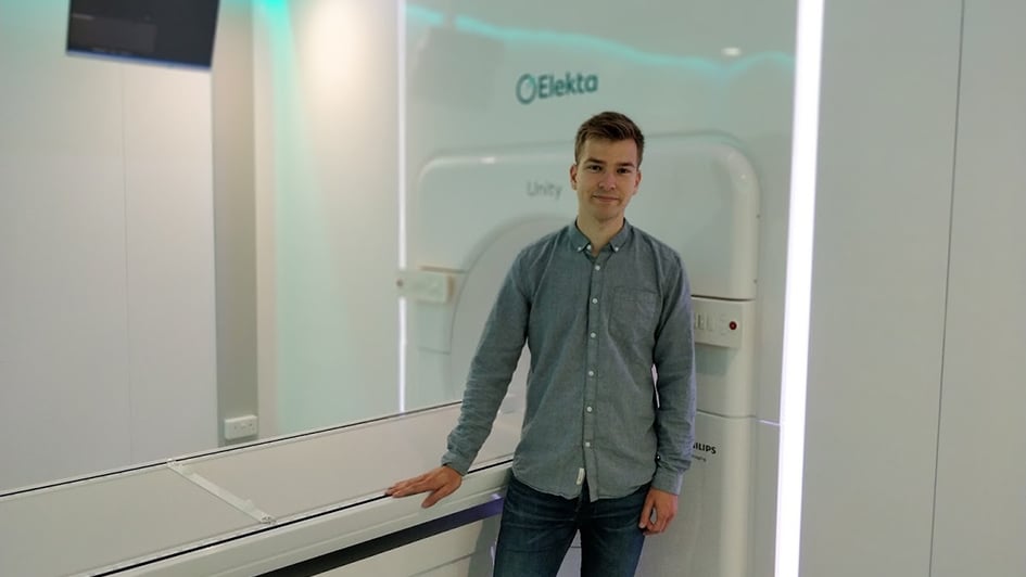
Joshua Freedman is a third-year PhD student in our Division of Radiotherapy and Imaging. In this blog post, he describes his research to help develop new approaches to support treatment planning and guidance on the MR Linac, a revolutionary new type of radiotherapy machine which is currently being applied for the first time in the UK on patients.

Image: PhD student Joshua Freedman standing in front of the MR Linac
Approximately 40 per cent of all cancer patients are treated with radiotherapy. It damages cancer cells and stops them from growing or spreading in the body.
In UK hospitals, healthcare professionals currently perform radiotherapy treatment in three main stages: treatment planning, treatment delivery and patient follow-up.
In the treatment planning stage, dosimetrists and physicists carefully design the radiation dose delivery – in terms of both the intensity of X-rays delivered to the tumour site and the shape of the radiation beam, which can be tailored to minimise radiation dose to healthy organs such as the heart, and maximise dose to the tumour sites.
Clinicians use CT (computed tomography) scans of each patient taken beforehand to plan treatment, and then carefully position the patients during treatment so that radiation can be delivered according to the plan.
The treatment course is usually delivered in small rounds of treatment (known as fractions) over a period of several weeks.
Currently, the same radiotherapy treatment plan is used for all fractions, but this approach has setbacks: a patient’s anatomy might undergo slow changes between treatment fractions, for example because of weight-loss or tumour shrinkage.
Another issue is that conventional radiotherapy is not able to fully account for any rapid anatomical changes during treatment delivery, for example, as a result of breathing, or due to bladder filling.
The ICR and The Royal Marsden have delivered the first ever treatment in the UK using a Magnetic Resonance Linear Accelerator (MR Linac) machine.
Real-time imaging
A revolutionary new type of radiotherapy machine – the MR Linac, which combines high-quality magnetic resonance (MR) imaging with radiation delivery – might deliver a solution to these challenges. I have the pleasure of working with the MR Linac for my PhD, which is about to enter its final year.
The MR Linac could potentially precisely locate tumours at the time of treatment, tailor the shape of X-ray beams in real time, and accurately deliver doses of radiation even to tumours that are moving, for example as a patient breathes.
Better-personalised treatment plans using the MR Linac will also help to account for anatomical changes between and during treatment fractions.
The first MR Linac in the UK was recently installed at our partner hospital, The Royal Marsden NHS Foundation Trust, and in a hugely exciting milestone – we have just treated the first patient, after several months of work to prepare and optimise the machine with the help of healthy volunteers who underwent scans to help us test and calibrate the equipment.
In my PhD, I am devising methods to calculate four-dimensional (volumetric space + time) MR imaging for the thorax, which might be employed on the MR Linac to better account for respiratory motion during radiation delivery, for instance in the treatment of lung cancer.
My journey to develop four-dimensional MRI began with an exciting ten-week summer internship in Professor Jeff Bamber’s Ultrasound team, where I was introduced to medical physics and worked on the photoacoustic system.
Shortly afterwards I re-joined the ICR to work with Professor Martin Leach and Professor Uwe Oelfke, in the Division of Radiotherapy and Imaging.
At the ICR we offer clinicians a variety of opportunities – from a taught master’s course in Oncology to fellowships providing protected time for research, and higher research degrees.
Studying at the ICR
I have really enjoyed studying at the ICR due to the immense variety of work on offer.
Some of the highlights of my time studying at the ICR include:
- Scanning volunteers and patients on the MR Linac and on diagnostic systems.
- Developing new methods to better visualise moving tissues for radiotherapy planning.
- Applying state-of-the-art Google software to image reconstruction problems.
- Writing journal articles.
- Attending various international conferences.
However, my favourite part of studying at the ICR has been working with a brilliant, friendly and supportive team. I have gained so many new skills from working in the Division of Radiotherapy and Imaging and am excited for my final year.
Original Source: https://www.icr.ac.uk/blogs/tales-from-the-lab/page-details/revolutionising-radiotherapy-with-the-mr-linac
Original Date: Sept 25 2018
Written By: Joshua Freedman