Patient rotation enables fixed-beam radiotherapy system

Prototype radiotherapy system
The prototype radiotherapy system combines a fixed vertical radiation beam with horizontal patient rotation. (Courtesy: Paul Liu)

Radiation therapy plays a fundamental role in cancer treatment, but there is a global shortage of radiotherapy centres, with many low-to-middle-income countries having limited or no treatment capability. This situation exists in part due to the cost of facilities and the expense of acquiring and operating radiotherapy systems. Linear accelerators with simplified designs, such as fixed gantry systems, could reduce these costs.

Researchers at the ACRF Image X Institute at the University of Sydney are developing a 3D conformal radiotherapy system with a fixed vertical X-ray beam, horizontal patient rotation and image guidance. The full-size proof-of-concept prototype, which offers high-quality radiation therapy from a smaller, more robust and more cost-effective system, has now been successfully commissioned (Med. Phys. 10.1002/mp.13356).

From a financial perspective, there are many potential advantages of a such a fixed-beam system. Without a rotating gantry, the system has fewer moving parts, which could improve reliability and robustness, and potentially reduce maintenance costs. It would also require less bunker shielding to operate safely, thereby reducing the cost of building new bunkers or renovating bunkers housing older radiotherapy equipment such as cobalt-60 units.

The prototype system — developed by Paul Liu and colleagues working on the Nano-X project to improve global access to radiotherapy — is based on the concept of patient rotation, specifically, keeping the radiation beam stationary while still achieving the necessary beam angles to achieve a desired dose distribution. Image guidance technologies will identify the tumour and adapt the treatment in real-time to ensure that the radiation dose is precisely delivered to the target.

The prototype comprises a standard Synergy linac with the gantry fixed at 0° and a horizontal patient rotation system (PRS). The PRS is a custom-designed radiotherapy couch equipped with straps for the head, chest, hips and legs, plus three independently controlled airbags that inflate over a patient’s chest and sides. The couch can move with two degrees-of-freedom to position and rotate the patient.

Patient rotation

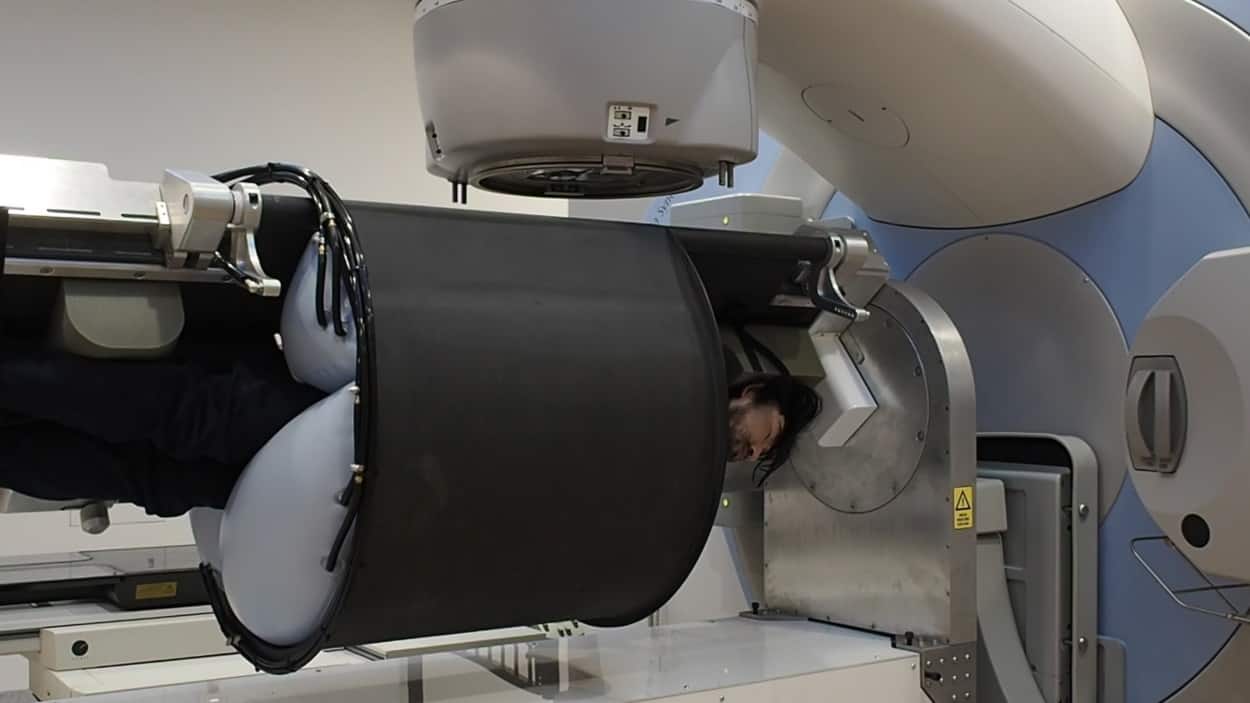
After the patient is immobilized and in a specified treatment position, they can be rotated to a specific angle for either kilovoltage (kV) imaging via the on-board imager or treatment with the megavoltage beam. The software operating the PRS allows for precise motion control, setting the target position or angle along with the desired velocity, acceleration and deceleration. It can also follow a series of queued motion commands, or execute quick-stop, return-to-home and patient egress commands.

The system passed all commissioning steps, which involved verification of geometric and dosimetric accuracy following conventional radiotherapy guidelines. The team also performed thorough testing of safety and interlock systems.

Clinical potential

The authors note that three essential steps will be needed before treating patients. Cone-beam CT image reconstruction under gravitational deformation may require advanced image reconstruction algorithms. They also need to develop methods to shift the beam to account for gravitational deformation-induced target motion.

Additionally, a patient’s tolerance of, and anxiety level relating to, horizontal rotation is unknown. It could be as much of a problem as an MRI exam is to a claustrophobic or noise-averse patient. An upcoming clinical trial will investigate and quantify how patients respond to strap and airbag immobilization and horizontal rotation.

Liu discussed the challenges with Physics World. “While we initially focused on static targets, an important part of the system will be its ability to adapt to motion, both from the patient’s normal physiological functions like breathing and from gravity as the patient is rotated,” he explains. “The next stage of the project will focus on implementing and testing algorithms that we’ve developed to both identify the amount of motion and to compensate for it accordingly.”

To enable real-time image guidance, the researchers are testing kilovoltage intrafraction monitoring (KIM), a novel tumour localization system developed at the University of Sydney that accurately estimates the 3D position of a target based on the 2D position of segmented markers in kV projections. Read more

Real-time image-guided ART achieved on a standard linac

“KIM will offer real-time 3D target tracking with sub-degree and sub-millimetre accuracy,” Liu says. “We have successfully tested KIM together with real-time multileaf collimation tracking on a miniature version of this system, and are currently scaling these algorithms to our full-size prototype. We will be using KIM with a deformable phantom where the target will move as it undergoes rotation.”

The researchers are also investigating intensity-modulated radiotherapy and volumetric-modulated arc therapy, which are under various stages of implementation. Liu says that both are technically feasible, because the software and hardware control of the PRS has sufficient precision and flexibility.

Much work, followed by testing with veterinary radiation treatments, will be required before the first palliative treatments on human cancer patients can be undertaken. The system is not designed for infants, very small children or obese patients. But for all other cancer patients, this prototype radiotherapy system has potential to fill the existing and expanding gap between available treatment and need, especially for patients living in economically challenged areas of the world.

Original source: https://physicsworld.com/a/patient-rotation-enables-fixed-beam-radiotherapy-system/

Original Date: Feb 14 2019

Original Author: Cynthia E Keen

Elekta: New Study to Learn From Every Cancer Patient Treated With Magnetic Resonance Radiation Therapy

The MOMENTUM study is a transformative approach to evaluating innovative medical technology

UTRECHT, The Netherlands, Feb. 4, 2019 /PRNewswire/ —

Today, the international MR-linac Consortium announced the launch of the MOMENTUM study. The study is designed to generate data that enable safe, fast and, above all, ‘evidence-based’ introduction of magnetic resonance radiation therapy (MR/RT) into clinical practice. The MOMENTUM study represents the next step in the development of the Elekta Unity MR/RT system; the study will be focused on building a robust body of real-world clinical evidence and insights made possible by this technology. Information gained through the MOMENTUM study will guide the use of MR/RT to improve outcomes for cancer patients.

“Each treatment session on this innovative system is an opportunity to gain insight into the benefits that this technology provides and, critically, to determine which patients benefit from MR/RT therapy,” said Dr. Helena Verkooijen, Professor of Evaluation of Innovation at University Medical Center Utrecht (UMCU) and a member of MOMENTUM’s Management team.

Radiotherapy is an important component many cancer treatment regimens and approximately 50% of all cancer patients receive radiation during their treatment journey*. As with most medical therapies for cancer, radiotherapy is associated with short- and long-term side effects that can be treatment-limiting and/or reduce patients’ quality of life during and after therapy. Many of these side effects result from radiation-related damage to healthy tissue. The MR-linac system is designed to address this challenge by allowing improved targeting of radiation to the tumor and reduced exposure of nearby tissues and organs.

Dr. William Hall, Assistant Professor of the Department of Radiation Oncology at the Medical College of Wisconsin noted. “We believe that this kind of rigorous and coordinated approach has tremendous potential to improve patient outcomes and change radiotherapy.”

Cancer centers participating in MOMENTUM will ask patients if they are willing to share de-identified information about their treatment and subsequent experience, including tumor control rates and quality of life. This information will be aggregated into repositories that will allow researchers to assess outcomes, enhance the product and evaluate alternative treatment approaches.

“The MR-linac Consortium includes some of the world’s most talented and dedicated cancer researchers,” said Dr. John Christodouleas, Vice President of Medical Affairs and Clinical Research at Elekta and a member of MOMENTUM’s management team. “By collaborating on the MOMENTUM Study, we expect to accelerate clinical innovations enabled by this breakthrough technology.”

Elekta Unity makes it possible to visualize the tumor with high-resolution images during treatment through combining high-field MRI technology with a linear accelerator. This allows extremely precise delivery of the radiation dose, enabling higher dosing to the tumor bed while better sparing the surrounding healthy tissues. While this is expected to lead to better tumor control and fewer side effects it is crucial to show that the advanced technical capabilities of MR/RT translate into real benefits for the patient, such as prolonged disease-free survival and better quality of life.

The innovative MR-linac technology was developed by Elekta in collaboration with the MR-linac Consortium, which comprises experts in oncology, radiation therapy, epidemiology and medical physics from leading cancer centers around the world.

Elekta Unity has CE-mark and 510(k) clearance but is not commercially available in all markets.

About the MR-Linac Consortium

The Elekta MR-linac Consortium is a collaborative industrial-academic partnership that Elekta founded with seven centers and our technology partner, Philips in 2012 to provide an evidence-based introduction of the MR-linac to the medical community, and to support the advancement of the technology. The institutions that participated are: (Founding members) University Medical Center Utrecht, the Netherlands; The Netherlands Cancer Institute-Antoni van Leeuwenhoek Hospital, the Netherlands; The University of Texas MD Anderson Cancer Center, USA; the Institute of Cancer Research, working with its clinical partner The Royal Marsden NHS Foundation Trust, UK; Froedtert & the Medical College of Wisconsin Clinical Cancer Center at Froedtert Hospital, USA; The Christie NHS Foundation Trust, UK; Odette Cancer Centre, Sunnybrook Health Sciences Centre, Canada. Lygature, The Netherlands, provides the public-private partnership management of the MOMENTUM study.

About Elekta

For almost five decades, Elekta has been a leader in precision radiation medicine. Our nearly 4,000 employees worldwide are committed to ensuring everyone in the world with cancer has access to – and benefits from – more precise, personalized radiotherapy treatments. Headquartered in Stockholm, Sweden, Elekta is listed on NASDAQ Stockholm Exchange. Visit elekta.com or follow @Elekta on Twitter.

Original Source: https://www.biospace.com/article/releases/elekta-new-study-to-learn-from-every-cancer-patient-treated-with-magnetic-resonance-radiation-therapy/

Original Source: https://www.biospace.com/article/releases/elekta-new-study-to-learn-from-every-cancer-patient-treated-with-magnetic-resonance-radiation-therapy/

Original Date: Feb 4 2019

Written By: Elekta

Answers to The Top 4 Questions About Radiation Therapy

Anyone that is starting a new medical treatment has a right to be a little nervous.  This article is being written to help ease the anxiety of cancer patients that are scheduled for radiation therapy.  Fear comes because of the unknown and common misunderstandings patients have about radiation. 

Radiation therapy is delivered using a large piece of medical equipment known as a linear accelerator.  With maintenance, repairs, and part replacement linear accelerators can deliver radiation to an average number of patients for between five and ten years.  Below we will answer the most commonly asked questions about radiation therapy using a linear accelerator, LINAC.

Is Radiation Therapy Painful?

Thankfully radiation therapy is not painful.  Patients often report that they do not experience any sensation when the radiation is delivered from the LINAC.   A few patients have reported that they feel a slight warm tingle in the area where the LINAC is delivering radiation.  The skin in the area where linear accelerators deliver treatment can become dry and itchy over time.  This can cause some discomfort but definitely not enough to stop treatment.  Skin reactions due to radiation can be treated with over the counter ointment. 

Does Radiation Therapy Cause Me to Be Radioactive?

Radiation therapy only makes patients radioactive when internal radiation is given.  Patients are radioactive while the radioactive materials are in them.  These patients are secluded in a private hospital room.  Patients that are treated using a LINAC through external radiation, will not be radioactive at all.  External radiation delivers a precise dose of radiation to the cancerous tissues instantaneously.  With external radiation the radiation does not linger. Once the LINAC is turned off the radiation isn’t an issue.  In external radiation, patients can continue on their normal routines without worry. 

Will I Lose My Hair During Radiation Therapy?

Radiation is considered a localized treatment which means that it focuses directly on the area being treated.  This being said you can expect hair loss in the area of treatment however unless treatment is done on your head you shouldn’t experience hair loss.  Confusion occurs because people associate radiation and chemotherapy as one in the same therapies.  The difference is that chemotherapy is a systemic treatment which means it affects the entire body.  During chemotherapy there is a likelihood that patients will lose their hair.

Should I Expect to Experience Nausea and Vomiting? 

Radiation therapy doesn’t usually cause patients to feel sick.  If treatment is given in areas such as the liver, brain, or gastrointestinal tract patients have more of a risk to experience nausea.  Also, patients that are going through chemotherapy and radiation at the same time there is more of a risk for feeling ill.

Radparts is the world’s largest independent distributor of OEM replacement parts for Linear Accelerators and Radiation Oncology equipment.  Radparts provides high quality, user friendly, low cost parts support for linear accelerators and radiation equipment. More information can be found at https://www.radparts.com/.


Does It Cost Money to Have an Old Linear Accelerator System Removed?


This is a frequently asked questions when healthcare centers are starting the process to budget for the purchase of a new or refurbished Linear Accelerator.  It is important to not be surprised when it comes to financial obligations within a medical organization.  The answer is Yes, there is a cost to have old LINAC systems removed from your facility.  There is a cost to have new equipment installed as well but that is often combined with the price you pay for the equipment itself. 

Although linear accelerators are a machine that offers a priceless treatment in the fight against cancer with lifesaving radiation beams treating thousands of patients for countless number of years, they do become worthless.  Like most machines, over time the labor and linear accelerator parts cost more to repair the LINAC machine than it is worth.  When this occurs, the machine is worth nothing and needs not only to be removed but also properly disposed of. 

 Another element to add to the mix is the introduction of new technology.  This is a reoccurring issue that is seen in medical equipment.  Older machines cannot be updated to run properly with the latest and greatest treatments and therefore simple become worthless.  Yep, even some LINAC machines with life left in them become obsolete to healthcare providers promoting the latest and greatest treatments.  Although these machines are worthless to these facilities they can often be moved and utilized by other facilities such as veterinarians, possible dermatologists, and of course in poverty-stricken locations that would otherwise go without. 

When a linear accelerator has been deemed to have little to know value, meaning no one is willing to pay you for it, your facility will face the cost of removal.  This cost includes professional and safe dismantling, removal, and disposal of the LINAC.  This process often requires the system to be loaded onto a crane as they weigh several thousand pounds.  This should be done by professionals to prevent catastrophe. After the machine has been removed from the facility it will be tested for radioactivity and disposed of once they are cleared. 

At the end of the day, when all is said and done the cost to have a large scaled piece of medical equipment such as a linear accelerator removed, dismantled, and disposed of ranges between $5,000 and $25,000.    

Radparts is the world’s largest independent distributor of OEM replacement parts for Linear Accelerators and Radiation Oncology equipment.  Radparts provides high quality, user friendly, low cost parts support for linear accelerators and radiation equipment. More information can be found at https://www.radparts.com/.


Elekta Unity MR-linac gains FDA 510(k) clearance

DOTmed.com – Elekta Unity MR-linac gains FDA 510(k) clearance The Elekta Unity magnetic resonance radiation therapy (MR/RT) system has gotten its FDA 510(k) premarket notification and is now ready for sale and clinical use in the U.S.

“Since receiving CE mark in June 2018, Elekta Unity has been transforming the care of cancer patients in Europe, and we are excited that this cutting-edge technology is now commercially available to U.S. patients,” Elekta president and CEO Richard Hausmann said in a statement, adding that the Unity will make possible the development of personalized, precision radiation therapy regimens optimized for safety and efficacy.

Special-Pricing Available on Medical Displays, Patient Monitors, Recorders, Printers, Media, Ultrasound Machines, and Cameras.This includes Top Brands such as SONY, BARCO, NDS, NEC, LG, EDAN, EIZO, ELO, FSN, PANASONIC, MITSUBISHI, OLYMPUS, & WIDE.

The Unity will “make radiation therapy a viable treatment option for more patients,” he added, thanking all involved in the MR-linac consortium and MR technology partner, Royal Philips.

The Unity is designed to simultaneously deliver radiation dose and visualization of tumors and adjacent healthy tissue in the form of high-quality MR images, employing integrated tools for possible treatment adjustments to match current anatomical information in a treatment session.

“Unity is a tremendous leap forward in our ability to tailor radiation therapy to each patient’s tumor and anatomy, and to adapt treatment in real time as the tumor changes shape and position relative to organs at risk,” said Dr. Christopher Schultz, chair of the Elekta MR-linac Consortium.

He called the new technology “fundamentally” transformational in terms of the development and implementation of therapy regimens that will permit clinicians “to achieve optimal outcomes for our patients.”

Back in September, Elekta forecast a net sales compound annual growth rate of 8-10 percent through its 2022/2023 financial year.

“We have improved our margin and cash flow and have returned to high growth. We are now in a good position to realize our vision,” said Hausmann in a statement, noting that “the future of our industry is in precision radiation medicine, including diagnostic quality imaging at the point of treatment, real-time adaptive treatment planning, data-driven personalization and intelligent automation.”

Its predictions follow the company’s recent decision to sell its magnetoencephalography (MEG) business to Croton Healthcare subsidiary York Instruments as part of an initiative to restructure and strategically prioritize its treatment solutions and oncology informatics portfolio, agreements set up between the radiotherapy manufacturer and other parties over the past year.

Original Source: https://www.dotmed.com/news/story/45569

Original Date: Dec 6 2018

Written By: Thomas Dworetzky

Trial results show compound makes radiotherapy more effective

A trial has shown that radiotherapy is more effective when levels of ropidoxuridine in a patients’ body reach a certain level…

radiotherapy

A new drug designed to make radiotherapy more effective in treating cancer has been given to patients while they are receiving radiation and shown to be safe.

The drug, called 5-iodo-2-pyrimidinone-2′-deoxribose (IPdR), or ropidoxuridine, has the advantage that patients can take it in capsule form, as opposed to intravenously. When the drug enters the body, researchers believe it changes into an active form that can make cancer cells more susceptible to the effects of radiotherapy.

Results of US NCI trial #9882, presented by Dr Timothy Kinsella from the Department of Radiation Oncology at the Warren Alpert Medical School of Brown University and Rhode Island Hospital in the USA, show that the drug has minimal side effects when given to patients with a variety of gastrointestinal cancers during the course of their radiotherapy.

Webinar: Improve ultrapure water system performance and uptime with ozone

Register for our on-demand webinar with SUEZ which is designed to help companies understand the application and benefit of ozone technology in ultrapure water systems and how ozone solutions can improve water system performance, uptime and product quality in pharmaceutical manufacturing applications.

Dr Kinsella explained: “The aim of my research is to find better ways to treat patients with cancer, and specifically to develop ways to make radiation treatment safer and more effective.

“Previous research found a promising compound called iododeoxyuridine, or IUdR, that worked very well to improve the effectiveness of radiotherapy, but IUdR could only be given intravenously and proved to have many side effects for patients.

“As a result, this new drug, IPdR, was developed. It’s a prodrug that can be taken as a capsule and, once inside the body, it’s converted into the active drug, IUdR.

“This trial is the first to test it out in patients while they are receiving radiation therapy, and the results suggest that it’s safe with minimal side effects.”

Dr Kinsella and his colleagues tested the new drug in a group of 18 patients with advanced cancers including oesophageal, pancreatic, liver, bile duct, rectal and anal cancers. All had been referred for palliative radiotherapy.

Alongside their radiotherapy, patients were given a daily dose of the IPdR prodrug over 28 days. They were given blood tests to check on the levels of both the IPdR prodrug and the active IUdR drug at various points during their treatment. The dose of the prodrug was gradually increased, and patients were monitored for side effects.

Results of the trial suggest that IPdR can be safely given to patients up to a dose of 1200mg per day for 28 days without causing serious side effects. The results also suggest that this dose creates levels of the active IUdR drug in patients’ blood that are high enough to have a radiosensitising effect.

Of the 18 patients on the trial, 14 could be assessed for any effect on their tumours with a CT or MRI scan 54 days after beginning the treatment. Among these patients, one had a complete response (disappearance of tumour), three showed a partial response (at least 30 percent reduction in the tumour targeted by radiotherapy), nine had stable diseases (no growth in the tumour) and one patient stopped treatment because of an infection and had progressive disease (at least 20 percent growth in the tumour).

Dr Kinsella added: “This clinical trial showed that when patients take IPdR at home before coming for radiation treatment, the level of IUdR in their bloodstream is high enough to make radiation more effective at killing cancer cells. It also showed that the dose of IPdR needed to achieve therapeutic levels of IUdR in the blood causes minimal side effects.

“However, this trial was with patients who had recurrent cancer and had already received a number of other cancer treatments. In newly diagnosed patients, it could be that we can safely use a higher dose and have a bigger effect on tumours.”

Dr Kinsella and his colleagues are already studying the effects of IPdR in patients receiving whole brain radiotherapy for cancer that has spread to the brain. Following this trial, plans are in progress to study the drug in patients who have been newly diagnosed with glioblastoma, an aggressive form of brain cancer.

Dr Eric Deutsch, Professor of Radiation Oncology and head of the radiation oncology department and research unit at the Institut Gustave Roussy, Villejuif, France, is a member of the EORTC-NCI-AACR Symposium scientific committee and was not involved with the research. He commented: “Radiotherapy is a vital element in treating many forms of cancer. This research is investigating whether the IPdR drug could make radiotherapy even more effective for more patients.

“In treating cancer patients, we must always consider the risks and benefits of any therapy. In this study, the risks of the IPdR drug were minimal, and the benefit was that it can be taken by patients at home. We don’t have enough evidence yet on whether IPdR can improve patient outcome, but we hope that this will become clearer as the research continues.”

The research was presented at the 30th EORTC-NCI-AACR Symposium on Molecular Targets and Cancer Therapeutics in Dublin, Ireland.

Original Source: https://www.europeanpharmaceuticalreview.com/news/81355/radiotherapy-more-effective/

Original Date: Nov 16 2018

Written By: European Pharmaceutical Review

Treating Cancer With Radiation Therapy


There are many parts and components that must be considered when servicing and repairing linear accelerators and other types of radiation therapy equipment.  Consider the inner workings of LINAC systems and the process they go through to deliver targeted radiation and you will see why professional servicing and maintenance is required.

External radiotherapy is done with the use of specialized radiation therapy equipment.  This equipment is designed to aim beams of radiation at the source of cancer.  The most common types of radiation being through the use of high energy x-ray beams. Other types can include particle beams, such as protons and electrons.  These beams are used to obliterate the cancerous cells within the are being treated while preventing radiation damage to healthy cells.

Radiotherapy works by harming the DNA within the cancerous cells.  The DNA is the genetic code which controls the behavior of the cells. Radiotherapy damages DNA directly on contact or creates charged up particles to damage the DNA.  This treatment should stop the growth or kill the cancer.  When cells die your body will break them down and get rid of waste substance.  Normal cells could be damaged but usually repair themselves. 

Before treatment can begin your doctor will want to go over the short- and long-term side effects. Most will be temporary and can be regulated with medication.  The team that treats you will use a combination of images including x-rays, CT scans, MRI scans, or PET scans.  They will be used to monitor the size of the tumor and measure the shrinkage that is occurring.

Radiotherapy machines, like linear accelerators, are very large and can look extremely intimidating. A LINAC uses electricity in creating radiotherapy beams.  The machine will never touch you and the radiation will not be felt.  Some discomfort can be expected from the side effects of treatment but can be controlled using medication. For radiotherapy to work the radiation must cover the entire cancerous area and the surrounding border.  Physicians will give the lowest dosage possible to prevent damage to the health tissue surrounding the cancer.  This will reduce the risks of side effects to the healthy tissue.

The dose of radiation you are prescribed will be divide up into small doses known as fractions. Instead of one large dose, these smaller doses allow the same amount of radiation overtime which helps to alleviate the side effects and allows the healthy tissues time between treatments to heal.  Radiation can be given as palliative care which is given to alleviate the pain associated with cancer or as a treatment to cure the cancer.

Radparts is the world’s largest independent distributor of OEM replacement parts for Linear Accelerators and Radiation Oncology equipment.  Radparts provides high quality, user friendly, low cost parts support for linear accelerators and radiation equipment. More information can be found at https://www.radparts.com/.


The World’s Largest and Most Powerful X-Ray Laser Just Went Online

The European XFEL is now online. Built to be the world’s largest facility to house the most powerful x-ray lasers that can be produced, the XFEL would contribute to a better understanding of molecular and chemical process by imaging particles.

Faster Imaging

The latest combined project by European nations is now online. It’s called the European X-Ray Free Electron Laser, or XFEL for short, and it’s now the largest and most powerful x-ray laser imaging instrument in the world, stretching some 3.4 kilometers (2.11 miles) long and 40 meters (11.15 feet) underneath the German city of Hamburg and a nearby town called Schenefeld.

The XFEL project began in 2007, with 11 nations partnering to make it a reality. After almost a decade of work and more than a billion euros spent, it’s now ready to contribute to the world of scientific research.

“It’s a fantastic and exciting day for us to open the European XFEL for operation after more than eight years of construction,” Robert Feidenhans’l, the facility’s managing director, said at Friday’s inauguration ceremony, the BBC reports. “I now declare we are ready to take data; we are ready to meet the challenge of getting groundbreaking results.”

x-ray laser european xfel particle imaging
Image credit: European XFEL

The superconducting linear accelerator housed in the underground tunnel is meant to run accelerated electrons to almost light-speed. These then pass through a slalom (or sloping) course of magnets called undulators where, as the electrons bend and turn, they emit flashes of X-rays. The XFEL generates these extremely bright and ultrashort pulses of light at a rate of up to 27,000 pulses per second, which is 200 times faster than other x-ray lasers — a billion times faster than light generated by synchrotrons.

Molecular Snapshots

The European XFEL’s primary purpose is to help study particulate matter. For example, it can be used to map the three-dimensional structures of biomolecules and other biological nanoparticles. The idea is to capture them during the process, as they occur, whilst these samples speed through the facility. The XFEL can deliver trillions (1,000,000,000,000) of X-ray photons in one pulse that lasts just about 50 femtoseconds (0.000,000,000,000,05 sec), for each of its 27,000 pulses per second.

Using an advanced camera called the Large Pixel Detector (LPD), installed as part of the XFEL by British engineers, it would allow researchers to capture molecular snapshots like never before. The cameras have a frame rate of 4.5MHz — 4.5 million pictures per second. Furthermore, each single image can be put together to create a “molecular movie” that details biochemical and chemical reactions in progress.

“The LPD captures the pattern of the X-rays after they’ve scattered through whatever it is the scientists have put in the beam. Its imaging surface is about half a meter by half a meter,” lead engineer Matthew Hart told the BBC. “It’s something we’ve been working on for 10 years, and it’s incredibly bespoke. It has to run really fast, handle really intense levels of X-rays but at the same time capture very small signals as well; and have very low noise.”

In a press release about the launch, Olaf Scholz, Hamburg city’s mayor had this to say: “With the European XFEL, scientists will forge ahead into unknown worlds and help to find answers to questions facing humanity that will make life on our planet better.” Analyzing atomic structures can, for instance, help in understanding various process involved in diseases. Indeed, the European XFEL could even become an important tool in exploring the world of quantum particles.

Original Source: https://futurism.com/the-worlds-largest-and-most-powerful-x-ray-laser-just-went-online/

Original Author: Dom Galeon

Original Date: Sept 5 2017