Early adopters are exploiting a novel motion phantom to explore the possibilities of real-time magnetic-resonance guided radiotherapy for improved cancer treatment and neurosurgery

Upcoming techniques based on magnetic-resonance guided radiotherapy (MRgRT) could enable clinicians to compensate for patient movements to a much higher degree, thanks to the clarity offered by MR imaging. Real-time tracking methods that can pinpoint changes in the position of a tumour translate to improvements in dose conformality by keeping radiation on target and sparing healthy tissue.
As vendors, early adopters and clinicians bring new ideas to fruition, a key part of their success depends on having the right development tools, which includes motion (or 4D) phantoms. Accurate models give researchers the chance to safely explore solutions for overcoming hurdles that can be faced in the clinic as a result of tumour motion. Scenarios include when a patient breathes, causing organs and tumours to move, or when there’s peristaltic motion through the digestive system.
We’ve designed our system to be compatible and expandable, and even – to a certain degree – customizable
Enzo Barberi, director of MR product development at Modus QA
Tumour movement has always challenged cancer treatment and manufacturers have worked hard to mitigate the issue as much as possible.
“Image guidance for radiation therapy has been around for well over a decade and most linacs have some form of cone-beam CT or EPID imaging that allows to them to roughly see where the target is,” says Enzo Barberi, director of MR product development at Modus QA – a developer and manufacturer of quality assurance tools for advanced radiotherapy and medical imaging. “But those imaging techniques provide little information about soft tissue.”
In contrast, MR imaging can reveal soft tissue in exquisite detail, which – when linked to a radiotherapy system – shines a welcome light on where the cancer is at any moment in time.
Barberi, who’s been working in this field for almost three decades, confirms that it’s a very exciting time in terms of the technology and the clinical development of next-generation techniques exploiting MR linacs. “In both systems that are available today, you can image while you are applying radiation,” he points out.
Real-time imaging hits the target
On-board MR imaging offers numerous possibilities for advancing radiotherapy treatment. For example, if gas happens to pass through the intestinal tract of a patient during radiation treatment, real-time MR imaging can detect whether the tumour has moved. And, if the target is now positioned outside the safety margins, the beam can be turned off until the gas has passed through and the tumour moves back into position.
“It’s a dramatic example of how the combination of these two techniques in parallel and in real-time can make a big difference in terms of accuracy in hitting the target when it’s moving,” Barberi comments. Real-time imaging using MR could also see the end of so-called gating, where patients are required to hold their breath to keep their chest stationary – a development that could speed up treatment as well as reducing discomfort.
Bringing these new techniques into the clinic requires reliable tools for quality assurance (QA). MRI-compatible models make it possible to test the ability of novel imaging sequences to track a wide range of movements – such as those resulting from respiration. Verification is important too.
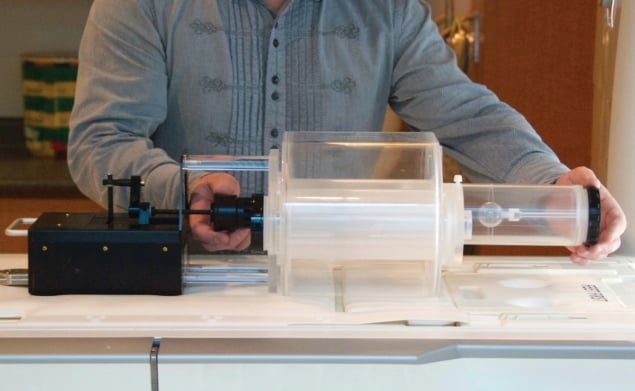
“Using phantoms like Modus’ programmable QUASAR MRI 4D motion product in combination with dosimetry inserts allows early adopters to calculate and measure the dose that is administered to a moving target and ensure that they are actually hitting this moving target and not the surrounding healthy tissue,” says Barberi.
These early adopters are important beta-testers for Barberi and his team, as they are at the frontier of MRgRT. Users require a phantom design that’s flexible, practical and easy to deploy, allowing them to gather as much data as possible for a range of possible patient scenarios.
“Modus focuses very heavily on workflow as we understand that time on the system is valuable,” Barberi comments. “If we can make our QA tools and QA procedures fast and efficient then sites are not only more likely to use them, but they will also appreciate the fact that we’re not taking up a lot of their magnet and linac time simply for setup or integration or when they have to switch over from one mode of measurement to another.”
Early adopters drive development
Features of the QUASAR motion phantom include a spherical target that can mimic numerous trajectories of a tumour in the body, including those seen during breathing. “We can add not only linear motion in and out of the phantom, but we can also add twist and offset that sphere so that it follows a complex 3D path as time plays out,” Barberi explains.
His team acknowledges that different investigators will have different demands, such as when it comes to dosimetry. “Users may wish to use ion chambers or film dosimetry or 3D gel dosimetry,” Barberi notes. “So we’ve designed our system to be compatible and expandable, and even – to a certain degree – customizable.”
Barberi’s group is already working on a second wave of inserts for the MR-safe motion phantom, thanks to the early-adopter programme. The new inserts will focus not just on soft tissue sites, but also modelling deep organ areas and more complex types of motion.

First UK radiation treatment using MR-guided linac
There is no shortage of challenges coming down the pipeline, but Barberi has a great team and is confident in Modus’ approach – having seen its flagship products develop successfully along a similar path. “Working with many different clinicians, physicists and OEMs over the years, we have families of different inserts that we can draw upon,” he says.
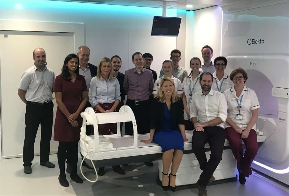
Barberi describes MRgRT as a “game changer”, and companies such as Modus are part of a big global effort to support upcoming advances that serve to accelerate the adoption of MR-linac systems for clinical treatment. Initiatives include STARLIT (System Technologies for Adaptive Real-time MR image-guided Therapies), a consortium developing techniques for next-generation motion compensation that includes two large equipment vendors – Elekta and Philips – along with small- and medium-sized companies and academic centres. “We are also equally proud to be a partner with ViewRay, supporting the requirements of an equally respected vendor, and their early adopting customers,” he says
Original Source: https://physicsworld.com/a/4d-phantom-eases-route-to-adaptive-mr-guided-radiotherapy/
Original Date: Sept 25 2018
Sponsored by Modus QA